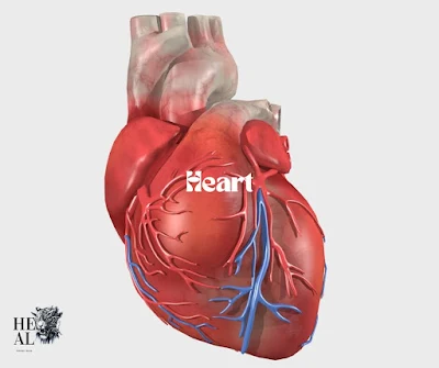
Heart
The heart is a powerful, hollow, cone-shaped muscular organ that sits at the center of your chest. It is enclosed within a protective layer called the pericardium and is primarily responsible for pumping blood throughout your body to meet its metabolic needs. As the core of your circulatory system, the heart ensures that every organ gets the oxygen and nutrients it needs. Without the constant work of this vital organ, your body wouldn’t be able to function properly.
 |
| heart |
Anatomical Position and Dimensions
- Positioned just behind the breastbone (sternum), with roughly one-third of the heart on the right side and two-thirds on the left.
- It measures approximately 12 x 8.5 x 6 cm.
- In males, the heart weighs around 310 grams, and in females, it weighs about 255 grams.
External Relations of the Heart
- Front: The heart is situated behind the body of the sternum, along with costal cartilages and the left lung.
- Back: Behind the heart, you’ll find the esophagus, thoracic aorta, and veins like the azygos and hemiazygos.
- Top: Near the top is the bifurcation of the pulmonary trunk.
- Bottom: The diaphragm supports the heart from below.
- Sides: It is flanked by both lungs and pleurae.
Layers of the Heart Wall
The heart consists of three layers:
- Epicardium: The outermost layer, also known as the visceral layer of the pericardium.
- Myocardium: The thick, muscular middle layer responsible for contracting and pumping blood.
- Endocardium: The innermost layer, composed of specialized cells that line the heart's chambers.
Structure and Chambers
The heart is divided into two halves—right and left—separated by a septum. Each half contains two chambers:
- Right Atrium
- Right Ventricle
- Left Atrium
- Left Ventricle
Blood Flow Through the Heart
The flow of blood through the heart follows a complex yet highly coordinated process:
- Venous blood enters the right atrium from the body via the superior and inferior vena cava.
- The right atrium pushes blood into the right ventricle through the tricuspid valve.
- The right ventricle pumps the blood to the lungs for oxygenation.
- Oxygenated blood returns to the left atrium.
- The left atrium moves blood into the left ventricle.
- The left ventricle pumps oxygen-rich blood into the aorta to supply the body.
Heart Valves
Valves ensure one-way blood flow, preventing backflow:
- Atrioventricular Valves: Tricuspid (right side), Mitral/Bicuspid (left side).
- Semilunar Valves: Aortic (left side), Pulmonary (right side).
Blood Supply to the Heart
Two main coronary arteries nourish the heart:
- Left Main Coronary Artery: Supplies around 80% of the blood to the heart muscle. It splits into the left anterior descending and circumflex arteries.
- Right Coronary Artery: Supplies the right ventricle, right atrium, and parts of the left ventricle.
Venous Drainage and Lymphatics
The heart's venous blood drains through the coronary veins into the coronary sinus, which empties into the right atrium. Lymphatic drainage is directed primarily to the brachiocephalic and tracheobronchial nodes.
Nerve Supply
The heart is regulated by the autonomic nervous system, which adjusts its function according to your body’s needs:
- Sympathetic Nervous System: Speeds up the heart rate.
- Parasympathetic Nervous System: Slows the heart rate.
- Conduction System: Includes key structures like the sinoatrial node and atrioventricular node, which generate and conduct electrical signals that trigger each heartbeat.
Maintaining Heart Health
- Balanced Diet: Eating a heart-healthy diet rich in fruits, vegetables, and whole grains is crucial for maintaining good cardiovascular function.
- Regular Exercise: Staying active helps the heart pump efficiently.
- Stress Management: Lowering stress levels can protect the heart and reduce the risk of heart disease.
- Avoid Smoking: Smoking can severely damage the heart and blood vessels, increasing the risk of heart attacks.
Conclusion
The heart plays a vital role in sustaining life by ensuring the constant flow of blood throughout the body. Its intricate design and function highlight its importance in keeping us healthy and alive. Understanding the heart, its chambers, valves, and blood flow processes can help us take better care of this essential organ.
